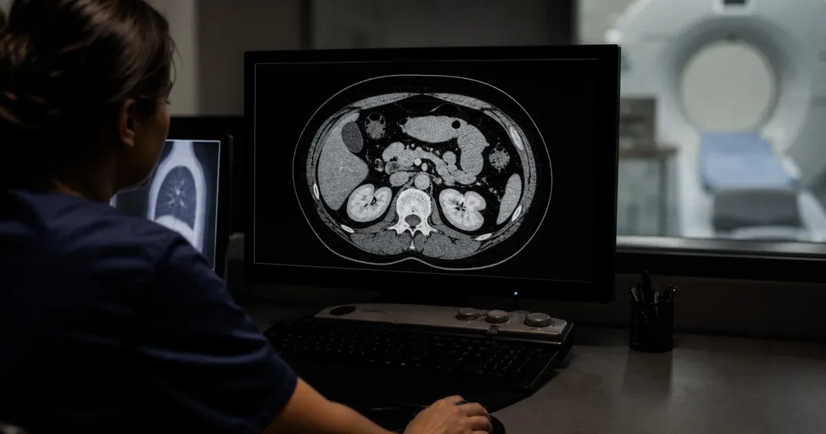
Introduction: Window Presets Are Starting Points, Not Absolutes
A CT scan stores far more grey-scale information than any monitor can display or the human eye can distinguish at once. Each voxel carries a Hounsfield Unit (HU) value on a scale that runs from roughly −1000 (air) through 0 (water) up past +1000 (dense cortical bone). To turn that wide numeric range into a viewable image, radiologists apply a window — a Window Width (WW) that sets how many HU are mapped across the full black-to-white ramp, and a Window Level (WL, also called window center) that sets the HU value sitting at mid-grey. This article is a practical, scannable reference of the standard CT window presets, expressed as WW/WL in HU, for brain, subdural, stroke, bone, lung, mediastinum/soft tissue, abdomen, liver, temporal bone, and vascular studies.
Before the table, one caveat that matters: these values are typical, approximate defaults, not fixed standards. Exact WW/WL presets vary between institutions, vendors, and individual radiologists, and modern PACS ship slightly different factory numbers. Treat every preset below as a sensible starting point that a reader fine-tunes at the workstation. For the mechanics of how WW and WL actually remap pixel intensities — and how DICOM stores default window values in the (0028,1050) Window Center and (0028,1051) Window Width tags — see our companion article, DICOM windowing: window width and level explained.
The HU Scale: The Anchors Every Preset Is Built On
Every window preset only makes sense relative to fixed points on the Hounsfield scale. These anchors are defined by the CT calibration itself and are worth committing to memory:
- Air: approximately −1000 HU (by definition).
- Fat: approximately −100 to −50 HU.
- Water: 0 HU (by definition).
- Simple fluid / CSF: roughly 0 to +15 HU.
- Soft tissue and most organs: roughly +20 to +60 HU.
- Acute blood / clot: roughly +50 to +90 HU.
- Contrast-enhanced vessels: often +100 to +300+ HU depending on phase.
- Cancellous (trabecular) bone: roughly +200 to +400 HU.
- Dense cortical bone: roughly +400 to +1000+ HU.
Because the diagnostic tissues in any given study cluster within a narrow band of this range, no single window can show everything. The whole point of a preset is to spend all the available grey levels on the HU band that matters for the task, letting everything outside the window clip to pure black or pure white.
Quick Reference: Common CT Window Presets (WW / WL)
The table below lists widely used presets. Values are typical approximations; your local defaults may differ by 10–20% or more.
| Window | Window Width (WW) | Window Level (WL) | Primary use |
|---|---|---|---|
| Brain (standard) | ~80 HU | ~40 HU | Grey/white matter differentiation |
| Subdural | ~130–300 HU | ~50–100 HU | Thin extra-axial collections near bone |
| Stroke / non-contrast brain | ~30–40 HU | ~35 HU | Early ischaemic grey/white loss |
| Soft tissue / mediastinum | ~350–400 HU | ~40–50 HU | Organs, muscle, mediastinal structures |
| Abdomen | ~350–400 HU | ~40–50 HU | Abdominal organs and fat planes |
| Liver | ~150 HU | ~30 HU | Subtle hepatic lesion conspicuity |
| Lung | ~1500 HU | ~−600 HU | Air-filled lung parenchyma |
| Bone | ~1500–2500 HU | ~300–500 HU | Cortical and trabecular bone detail |
| Temporal bone | ~4000 HU | ~700 HU | Ossicles, otic capsule, fine detail |
| CT angiography / vascular | ~700 HU | ~80 HU | Contrast-opacified vessels |
You can explore how these presets behave on real image data with our browser-based DICOM image viewer, and read the stored default window values from the header using the DICOM tag viewer. Everything runs client-side; no image ever leaves your machine.
Brain Window (WW ~80 / WL ~40): Seeing Grey vs White Matter
The standard brain window is deliberately narrow. Normal brain parenchyma spans only a small band — grey matter sits around +37 to +45 HU and white matter around +20 to +30 HU. That difference of roughly 8–12 HU is the entire signal a radiologist needs to detect oedema, mass effect, or early infarct. A width of about 80 HU centred near 40 HU spreads the full black-to-white ramp across just that band, maximising contrast between the two tissue types. Widen the window and the grey/white distinction flattens into uniform grey; the trade-off is that a narrow window makes the image visibly noisier, which is expected and acceptable at the head.
Stroke / Non-Contrast Brain (Narrow WW ~30–40 / WL ~35)
In suspected acute ischaemic stroke, radiologists often narrow the brain window even further — sometimes to a width of 30–40 HU with a level around 30–35 HU — to accentuate the loss of grey/white differentiation that marks early infarction. This so-called “stroke window” exaggerates a 2–4 HU drop in an ischaemic cortical ribbon that would be invisible on a routine brain window. It trades away almost all tolerance for noise in exchange for maximal sensitivity to subtle hypodensity. It is a refinement of the standard brain window, not a separate calibration — the same pixels, viewed through a tighter aperture.
Subdural Window (WW ~130–300 / WL ~50–100)
A thin subdural haematoma layered against the inner table of the skull can hide on a standard brain window because the bright bone and the bright blood merge visually. Widening the window to roughly 130–300 HU and raising the level toward 50–100 HU separates the acute blood (around +50 to +90 HU) from the adjacent cortical bone (hundreds of HU) so the collection becomes conspicuous against the skull. Many reading protocols mandate that every head CT be reviewed on brain, subdural, and bone windows precisely so that thin extra-axial collections near bone are not missed on brain settings alone.
Soft Tissue / Mediastinum & Abdomen (WW ~350–400 / WL ~40–50)
Soft-tissue windows are the general-purpose body setting. Muscle, solid organs, lymph nodes, and fat all fall within roughly −100 to +100 HU, so a width of about 350–400 HU centred near 40–50 HU shows organ margins, fat planes, and fluid collections together with usable contrast. The same approximate preset serves the mediastinum on chest CT and most of the abdomen, which is why a single “soft tissue” or “body” window covers a large share of routine reading. It is broad enough to keep both fat and enhancing tissue on-screen without clipping.
Liver Window (WW ~150 / WL ~30): A Narrowed Soft-Tissue View
Detecting a subtle hypo- or hyper-enhancing liver lesion demands more contrast than a general body window provides. Normal liver parenchyma enhances to roughly +100 to +120 HU in the portal-venous phase, and a lesion may differ from background by only 10–20 HU. Narrowing the window to about 150 HU with a level near 30–40 HU (some sites centre higher, near the enhanced parenchyma) amplifies those small HU differences so a lesion that is nearly invisible on the standard abdomen window stands out. It is the same principle as the stroke window — tighten the aperture over the HU band where the lesion lives.
Lung Window (WW ~1500 / WL ~−600): The Opposite Extreme
Lung parenchyma is mostly air, so the diagnostic HU band sits far down the negative side of the scale, from about −1000 HU (air) up to around −500 HU. A lung window uses a very wide width (1500 HU) centred deep in the negatives (−600 HU) so that vessels, bronchial walls, nodules, and the texture of interstitial disease are all visible against aerated lung. The width is large because lung findings span a broad HU range; the deeply negative level is what shifts the whole visible band into the air-and-vessel territory that soft-tissue windows clip to black.
Bone Window (WW ~1500–2500 / WL ~300–500)
Bone spans the widest useful HU range of any tissue — trabecular bone around +200 to +400 HU, dense cortical bone well above +1000 HU. A wide window (~1500–2500 HU) with a high level (~300–500 HU) maps that broad, high range across the grey ramp so cortical margins, trabecular pattern, and fracture lines are sharp. Bone windows are usually reconstructed with a sharp (high-frequency) kernel as well; windowing and reconstruction kernel work together, but they are independent settings — the window controls contrast display while the kernel controls spatial sharpness.
Temporal Bone Window (WW ~4000 / WL ~700): Maximum Width
The temporal bone contains the smallest, densest anatomy in routine CT — the ossicles, the otic capsule, fine bony canals. Resolving them demands an extremely wide window, around 4000 HU centred near 700 HU, so that the enormous HU contrast between dense petrous bone and the tiny air spaces of the middle ear is compressed onto the display without blooming. This is the widest window in common use, and it is paired with a very sharp reconstruction kernel and thin slices. Ordinary bone windows are too narrow to separate the finest ossicular detail.
CT Angiography / Vascular Window (WW ~700 / WL ~80)
Contrast-opacified vessels enhance strongly, often into the +200 to +400 HU range or higher at peak. A vascular window (~700 HU width, ~80 HU level) is shifted brighter and somewhat wider than a soft-tissue window so that the opacified lumen, vessel wall, calcified plaque, and any endoleak or filling defect are all distinguishable at once. Radiologists frequently adjust this on the fly per patient, since contrast timing and cardiac output shift the actual luminal HU from case to case — a vivid reminder that presets are starting points.
Why Presets Are Only a Starting Point
Every value in this reference is a convention, not a rule baked into the standard. Several factors push the optimal window away from the textbook default in any given study:
- Reconstruction kernel: sharper kernels add noise, sometimes prompting a slightly wider window.
- Contrast phase and timing: arterial vs portal-venous phases change luminal and parenchymal HU substantially.
- Patient body habitus and dose: higher noise at low dose can justify a wider window.
- Vendor and PACS defaults: factory presets differ between manufacturers, and the DICOM header may carry its own Window Center/Width the modality chose.
- Reader preference and pathology: the target finding dictates the tuning — a subtle liver lesion and a large abscess are not read at the same window.
This is why radiologists window interactively with the mouse rather than clicking a fixed preset and stopping. The presets get you into the right neighbourhood; the fine adjustment finds the finding. When a study opens with an odd default appearance, the DICOM header often explains it — you can read the stored (0028,1050) Window Center and (0028,1051) Window Width values directly with our DICOM tag viewer, and experiment with live windowing in the DICOM image viewer.
Conclusion
CT window presets exist because a single grey-scale ramp cannot show air, fat, fluid, blood, soft tissue, and bone all at once — each tissue lives in its own band of the Hounsfield scale, and a window is how you spend the display’s limited contrast where it counts. Learn the anchors (air −1000, water 0, fat ~−100, cortical bone +400 to +1000+), keep the typical WW/WL presets in this reference handy, and remember they are approximate, institution-dependent starting points to fine-tune, never fixed numbers to trust blindly. For the underlying mechanics of how WW and WL remap pixel values, continue with our companion piece on DICOM windowing explained, and for how those default values are encoded in the file header, see our DICOM value representations reference.
Sources
- Radiopaedia — Windowing (CT) — general reference for the WW/WL mechanic and typical window presets; exact values are institution-dependent.
- DICOM PS3.3 §C.11.2 VOI LUT Module — defines the Window Center (0028,1050) and Window Width (0028,1051) attributes stored in the image header.