Introduction: The Negative That Should Not Be There
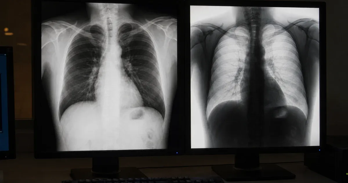
One of the most common support tickets in any imaging shop reads something like this: "The chest X-ray opens fine in our PACS, but when I load the same file in a different viewer the bones come out black and the lungs come out white — it looks like a photographic negative." The pixels are not corrupt, the transfer syntax decoded correctly, and the windowing values are sensible. What changed is the interpretation of a single short DICOM attribute: Photometric Interpretation, tag (0028,0004). This article explains exactly why DICOM images sometimes display inverted, how the MONOCHROME1 and MONOCHROME2 values differ, and where in the display pipeline the grayscale ramp gets flipped. You can inspect the relevant tags yourself with our DICOM Tag Viewer and confirm how a compliant renderer behaves in the DICOM Image Viewer.
What Photometric Interpretation Actually Declares
Photometric Interpretation is defined in the DICOM Standard, PS3.3 Section C.7.6.3.1.2 ("Photometric Interpretation"), as part of the Image Pixel Module. It is an enumerated string attribute with a Value Representation of CS (Code String). Its job is to tell the rendering software how to translate stored pixel sample values into a visible intensity or color. It is not a cosmetic hint — it is a normative instruction. A viewer that ignores it is non-conformant, not merely opinionated.
For grayscale images the standard defines two enumerated values that are mirror images of each other:
- MONOCHROME2 — the minimum stored pixel value is displayed as black and the maximum is displayed as white. Higher value means brighter. This is the convention nearly everyone intuitively expects.
- MONOCHROME1 — the minimum stored pixel value is displayed as white and the maximum is displayed as black. Higher value means darker. This is the inverse ramp.
Crucially, the underlying pixel data (7FE0,0010) can be byte-for-byte identical between two files; only the value of (0028,0004) determines whether the result looks positive or negative on screen. If a viewer hard-codes a "min equals black" assumption and ignores the tag, every MONOCHROME1 image it touches will render as a negative. That single oversight is the root cause behind the overwhelming majority of "inverted DICOM" reports.
Why MONOCHROME1 Exists at All
The MONOCHROME1 convention is a direct inheritance from analog film radiography. On exposed and developed film, areas that received more X-ray photons become darker (higher optical density), while areas shielded by dense bone stay light. Early digital systems — particularly computed radiography (CR) readers and film digitizers — mapped their stored values to mimic that film appearance, where larger acquired values corresponded to darker output. To preserve the look radiologists were trained on, those devices tagged their output as MONOCHROME1. DICOM did not invent the inversion; it merely gave the existing film-era polarity a formal name so that downstream software could reproduce it faithfully. Many modern digital radiography (DR/DX) units, by contrast, emit MONOCHROME2 because the calibration was redesigned around the "more signal equals brighter" model used by CT and MR.
Where in the Pipeline the Inversion Happens
DICOM specifies a well-ordered grayscale display pipeline in PS3.4 Annex N (the Grayscale Softcopy Presentation State) and in PS3.3 C.11. Conceptually, stored pixel values flow through a chain of transforms before they reach the monitor. The stages relevant to inversion are:
- Modality LUT — converts stored values into meaningful physical units (for CT, Hounsfield Units via Rescale Slope
(0028,1053)and Rescale Intercept(0028,1052)). - VOI LUT / windowing — applies Window Center
(0028,1050)and Window Width(0028,1051)to select the visible range. - Presentation LUT — the final mapping to "P-Values" (perceptually linear display values), governed by the Presentation LUT Shape
(2050,0020)attribute, which can take the valueIDENTITYorINVERSE.
Photometric Interpretation acts at the very start of this chain: it establishes the polarity convention of the stored values. A correct renderer treats MONOCHROME1 as if an inversion is baked into the data, so it must flip the grayscale ramp to produce a normal-looking image. The cleanest implementation detail is to apply the windowing math identically for both interpretations and then, only for MONOCHROME1, compute the final 8-bit display value as 255 - windowed. That one subtraction is the entire difference between a correct image and a negative.
Presentation LUT Shape Adds a Second Inversion Switch
To complicate matters, a Grayscale Softcopy Presentation State (GSPS) object or the Presentation LUT Shape attribute can independently request INVERSE polarity. This is what a "Invert" button in a PACS toolbar typically toggles. The standard is explicit that these transforms compose: if an image is MONOCHROME1 and a presentation state requests an additional inversion, the two effectively cancel, and you see a positive image again. A frequent debugging mistake is to apply both the MONOCHROME1 inversion and a UI-level invert, producing a double negative that looks correct on one file and wrong on the next. When you compare two viewers and the polarity disagrees, the question to ask is always: which layers of inversion is each one applying? Inspecting (0028,0004) and (2050,0020) side by side in the DICOM Tag Viewer usually resolves the argument in seconds.
How to Diagnose an Inverted Image
When a study renders as a negative, work through this checklist before assuming the file is broken:
- Read the tag. Open the file and look at Photometric Interpretation
(0028,0004). If it saysMONOCHROME1, an inversion is expected and your renderer must honor it. If it saysMONOCHROME2but the image looks inverted, the renderer is wrong, not the file. - Check Pixel Representation. Tag
(0028,0103)declares signed (1) or unsigned (0) data. A viewer that treats signed two's-complement CT data as unsigned will wrap negative Hounsfield values to huge positives, which can look like a partial inversion or "solarized" artifact rather than a clean negative. - Look for a Presentation LUT Shape. Tag
(2050,0020)set toINVERSErequests an additional flip. Confirm whether a presentation state object accompanies the image. - Verify the windowing tags. A negative Window Width is illegal under PS3.3 C.11.2 and, if mishandled, can also reverse contrast. Window Center and Window Width should be positive, sensible numbers for the modality.
- Test against a second conformant viewer. If a known-good viewer renders the file correctly and yours does not, the defect is in your rendering code, not the data.
A Concrete Worked Example
Suppose a CR chest image stores 12-bit unsigned values, with Photometric Interpretation set to MONOCHROME1, Window Center 2048, and Window Width 4096. A naive renderer computes the windowed value for a bright bone region as, say, 230 out of 255 and paints it light gray — but because the interpretation is MONOCHROME1, the conformant result should be 255 - 230 = 25, a dark pixel. Skip that subtraction and the bone glows white while the lung fields turn dark: the textbook inverted negative. The fix is not to change the window values, re-export the file, or "convert" anything; it is simply to apply the one-line inversion the tag has been requesting all along.
Should You Ever Rewrite MONOCHROME1 to MONOCHROME2?
It is technically possible to normalize a file by inverting the pixel data, setting (0028,0004) to MONOCHROME2, and saving. Some archives do this to simplify downstream tooling. However, the DICOM Standard treats pixel data as clinically authoritative, and modifying it has consequences. Any embedded VOI LUT, presentation state, or overlay that assumed the original polarity may now be inconsistent. For regulated workflows, the safer path is to keep the original pixels intact and fix the renderer so it honors the tag, exactly as PS3.3 requires. If you must transcode, record the change in the image's derivation description and treat the result as a derived object, not the original. De-identification and format conversion pipelines should be especially careful here, because re-encoding polarity silently can mask whether a viewer downstream is conformant.
Color and Multi-Sample Interpretations for Context
For completeness, Photometric Interpretation also governs color images, and those values never invert in the grayscale sense. RGB maps three samples directly to red, green, and blue channels; PALETTE COLOR uses indexed lookup tables defined in tags (0028,1101) through (0028,1203); and YBR_FULL, YBR_FULL_422, YBR_ICT, and YBR_RCT describe luminance-chrominance encodings used with JPEG and JPEG 2000 compression, defined in PS3.5. None of these carry the positive/negative polarity question, which is unique to the two monochrome variants. When an RGB endoscopy or dermatology image looks wrong, the cause is almost always a color-space or planar-configuration mismatch, not a MONOCHROME inversion.
Practical Takeaways for Imaging Teams
Inverted DICOM display is almost never a data-corruption problem and almost always an interpretation problem. The fix lives in three short attributes and one subtraction:
- Always read Photometric Interpretation
(0028,0004)and honor MONOCHROME1 by inverting the grayscale ramp. - Compose — do not stack blindly — the image-level inversion with any Presentation LUT Shape
(2050,0020)or GSPS-requested inversion. - Respect Pixel Representation
(0028,0103)so signed CT data does not wrap and masquerade as an inversion.
If you want to deepen your understanding of how the surrounding transforms interact, our companion guides on how DICOM image rendering works and standard CT, MRI, and X-ray windowing presets walk through the full pixel-to-screen pipeline in detail. To verify any of the claims here against a real file, drop a study into the DICOM Image Viewer and watch the polarity behave exactly as the tag instructs — entirely in your browser, with no pixel data ever leaving your device.
Conclusion
The next time a radiograph opens as a black-and-white negative, resist the urge to blame the file. Open it, read (0028,0004), and the answer is usually staring back at you: MONOCHROME1. The DICOM Standard, from PS3.3 C.7.6.3.1.2 through the presentation pipeline of PS3.4 Annex N, defines polarity precisely so that any conformant viewer can reproduce the intended appearance. Understanding that single attribute — and where in the rendering chain it acts — turns a baffling negative image into a one-line fix and a teachable moment for the whole imaging team.