Introduction: One Image, Many Appearances
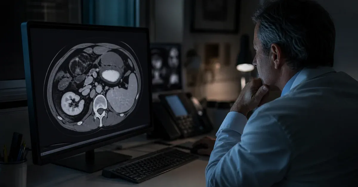
Open the same CT slice in two different presets and it can look like two different scans. Under one setting the lungs are crisp and black while the mediastinum is a featureless white blob; under another the soft tissues show subtle grey gradients but the lungs disappear into a uniform dark field. Nothing about the underlying data changed. What changed is the window — the mapping from stored pixel values to the shades of grey your monitor can actually display. This mapping is controlled by two DICOM attributes, Window Center and Window Width, often abbreviated WC/WW or window level / window width (WL/WW).
Windowing is one of the most fundamental concepts in diagnostic imaging, yet it is easy to use for years without understanding the numbers behind it. This article explains what windowing is, how it relates to Hounsfield Units in CT, exactly how the DICOM Window Center (0028,1050) and Window Width (0028,1051) attributes define what you see, and how linear windowing differs from a VOI LUT. You can read these values from any file with our DICOM tag viewer and see them applied live in the DICOM image viewer.
What Windowing Actually Is
A CT scanner records far more tonal detail than a display can show or a human eye can distinguish. A typical CT image stores 12 bits of precision per pixel, giving roughly 4,096 distinct values; on the Hounsfield scale these span from about −1,000 for air to well over +1,000 for dense bone and metal. A standard monitor, by contrast, presents only 256 shades of grey, and the eye can reliably separate far fewer than that in a single glance.
Windowing bridges that gap. Instead of squeezing the entire stored range into 256 greys — which would waste almost all of the display's contrast on tissues you do not care about — you select a subset of the value range and map only that subset across the full black-to-white ramp. Values below the window are clipped to pure black; values above it are clipped to pure white; values inside the window are spread linearly across the greyscale. By choosing which slice of the data to expand, you trade away contrast in the regions you are ignoring to gain contrast in the region you are examining.
Hounsfield Units: The Scale Being Windowed (CT)
In CT, the values being windowed are usually Hounsfield Units (HU), a calibrated scale of radiodensity. The scale is anchored at two fixed points: distilled water is defined as 0 HU and air as approximately −1,000 HU. Everything else falls relative to those anchors. Approximate values worth remembering:
- Air: about −1,000 HU
- Fat: roughly −100 to −50 HU
- Water: 0 HU (by definition)
- Soft tissue / muscle: roughly +30 to +80 HU
- Cancellous and cortical bone: from a few hundred HU up past +1,000 HU
Because the HU scale is standardized, a window expressed in HU means the same thing across scanners and sites, which is why CT presets like “soft tissue” or “lung” are portable. Note that HU is a CT concept; MR, ultrasound, and plain radiography also use windowing, but their pixel values are not Hounsfield Units and carry no absolute physical calibration.
From Stored Pixels to Hounsfield Units: Rescale Slope and Intercept
There is an important subtlety: the raw values in the Pixel Data element are usually not Hounsfield Units directly. They are stored integers that must first be converted using two more DICOM attributes, Rescale Intercept (0028,1052) and Rescale Slope (0028,1053). The relationship is a simple linear transform:
HU = (stored pixel value × Rescale Slope) + Rescale InterceptFor most CT data the slope is 1 and the intercept is −1,024 (or −1,000), which shifts the unsigned stored values down onto the signed HU scale. Windowing is then applied in the output units of that rescale — that is, in Hounsfield Units. When you debug an image that looks too bright or too dark, always check the rescale attributes first: a missing or wrong intercept throws the whole window off by a fixed offset.
The Two Attributes: Window Center and Window Width
DICOM defines the window with two attributes in the VOI LUT Module of PS3.3:
- Window Center
(0028,1050)— the midpoint of the window, in rescaled (HU) units. This controls brightness. Raising the center shifts the whole visible band toward higher values, which makes the image appear darker overall because more of the low-value tissue falls below the window; lowering it brightens the image. - Window Width
(0028,1051)— the total span of values the window covers, again in HU. This controls contrast. A narrow width spreads a small value range across the full greyscale, producing high contrast where small density differences become visible; a wide width compresses a large range into the same greys, lowering contrast but showing more tissues at once.
Both attributes use the DICOM VR DS (Decimal String), so they are stored as text and may carry multiple values (a file can offer several preset windows). A companion attribute, Window Center & Width Explanation (0028,1055), may hold human-readable labels such as “LUNG” or “BONE” for each preset.
How the Displayed Range Is Computed
From the center C and width W, the window's lower and upper bounds follow directly:
lowest displayed value  = C − (W / 2)
highest displayed value = C + (W / 2)
Every value at or below the lower bound renders as black; every value at or above the upper bound renders as white; values between them map linearly onto the greyscale. This is the classic linear windowing behavior, and the DICOM standard specifies the exact rendering function (including how the endpoints are handled) in the VOI LUT Module.
A Concrete Numeric Example
Take a common soft-tissue setting of C = 40, W = 400. The displayed range is:
low  = 40 − (400 / 2) = 40 − 200 = −160 HU  → black
high = 40 + (400 / 2) = 40 + 200 =  240 HU  → white
So every voxel at −160 HU or below appears pure black, everything at +240 HU or above appears pure white, and the range from −160 to +240 HU — which brackets fat, fluid, muscle, and organ parenchyma — is spread across the greys. Bone at +600 HU is off the top of the window and shows as flat white, while aerated lung at −800 HU is off the bottom and shows as flat black. That is exactly why bone and lung detail vanish at a soft-tissue window: they are being clipped.
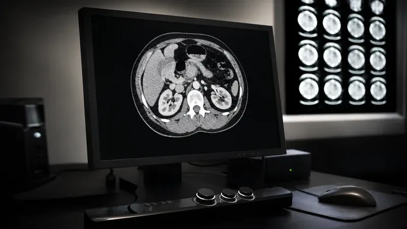
Now widen to C = −600, W = 1500 (a lung setting) and the range becomes −1,350 to +150 HU. Suddenly the aerated lung parenchyma occupies most of the greyscale and its fine architecture becomes visible, while the soft tissues collapse toward white. Same pixels, different window, entirely different diagnostic picture.
Why the Same Image Looks Different
This clipping-and-stretching behavior is the whole reason a single dataset supports many clinical “looks.” The pixel values never change; only the two numbers that define which slice of the value range gets the display's contrast do. Radiologists routinely cycle through several windows on the same series — soft tissue, then lung, then bone — because each one reveals structures the others hide. Presets are simply named pairs of C and W chosen for a given tissue and task; we cover the common CT presets in detail in our companion guide on CT window presets for brain, lung, bone, and soft tissue.
An important consequence for anyone building or configuring viewers: the Window Center and Width stored in a file are only a suggested default chosen by the modality or a technologist. They are a starting point, not a fixed property of the image. Any viewer is free to override them, and clinicians adjust them interactively at the workstation on nearly every study.
Linear Windowing vs VOI LUT
The center/width pair describes a linear Value of Interest transform: a straight-line ramp between the two bounds. DICOM also allows a more general, non-linear mapping through an explicit VOI LUT (Value of Interest Look-Up Table), defined by the VOI LUT Sequence (0028,3010) in the same VOI LUT Module of PS3.3. Instead of a formula, a VOI LUT provides an explicit table that maps each input value to an output display value, allowing arbitrary curves — useful when a simple linear ramp cannot render the intended tonal response.
When both are present, the VOI LUT Sequence takes precedence over the Window Center/Width values, though many CT workflows only ever use the linear window. There is also an attribute, VOI LUT Function (0028,1056), that can select a SIGMOID response instead of the default LINEAR one when using center/width. For the full authoritative definition, the VOI LUT Module in DICOM PS3.3 is the reference; the practical takeaway is that “windowing” in everyday CT almost always means the linear center/width transform described above.
Reading Window Values From a Real File
To see the actual windowing metadata a modality wrote, open the study in our browser-based DICOM tag viewer and look for (0028,1050) Window Center and (0028,1051) Window Width, along with (0028,1052) Rescale Intercept and (0028,1053) Rescale Slope. Everything runs locally in your browser — no file ever leaves your machine. If you want to watch different window settings change the appearance interactively, load the same file into the DICOM image viewer. For a broader tour of DICOM metadata types, see our DICOM value representations reference, and for how these attributes are encoded on the wire, our guide to DICOM transfer syntaxes.
Conclusion
Windowing is the deliberate act of throwing away contrast you do not need so you can see the contrast you do. Two DICOM attributes carry the whole decision: Window Center (0028,1050) sets brightness by choosing the midpoint, and Window Width (0028,1051) sets contrast by choosing the span, with the displayed range running from C − W/2 to C + W/2. In CT those numbers live on the calibrated Hounsfield scale after the rescale slope and intercept convert the stored pixels, which is what makes presets portable across scanners. Understand these few numbers and the puzzling variability of medical images resolves into something predictable and controllable — a single dataset you can interrogate from many angles by changing nothing but the window.
Sources
- DICOM PS3.3 §C.11.2 — VOI LUT Module — normative definition of Window Center (0028,1050), Window Width (0028,1051), the VOI LUT Sequence, and the linear/sigmoid rendering functions.
- DICOM PS3.3 — Information Object Definitions — the full standard part covering attribute modules, including rescale and pixel presentation.
- Radiopaedia — Windowing (CT) — general clinical reference on CT windowing, Hounsfield Units, and common tissue presets.